Bones of the SKULL - LEARN IN 4 MINUTES | 3D Anatomy of Skull Bone | Skull Bones - Lecture #3
#anatomy #skull #skullanatomy
In this YouTube video we embark anatomy of the skull bone, which forms the protective enclosure for our brain and provides structure and support for our facial features. Delve into the intricate network of bones, sutures, and foramina that make up the skull, understanding their functions and interconnections.
Through vivid animations and cutting-edge 3D visuals, we bring the intricate details of the skull bone to life, highlighting its various regions and landmarks.
The human skull is composed of several cranial bones that protect and encase the brain. The names of these cranial bones are as follows:
1-Frontal bone: Forms the forehead and the upper part of the eye sockets (orbits).
2-Parietal bones (right and left): Form the sides and roof of the cranium.
3-Temporal bones (right and left): Located on the sides and base of the skull; house the ears and jaw joint (temporomandibular joint).
4-Occipital bone: Forms the back and base of the skull.
5-Sphenoid bone: Sits at the base of the skull, contributing to the floor of the cranium and housing important structures like the pituitary gland.
6-Ethmoid bone: Located in front of the sphenoid bone, it contributes to the nasal cavity and the orbits.
Here are the main facial bones:
1-Maxilla (right and left): These are the upper jawbones that form the central portion of the face, supporting the upper teeth and contributing to the structure of the nasal cavity, orbits, and hard palate.
2-Mandible: Commonly known as the jawbone, it is the largest and strongest facial bone. The mandible holds the lower teeth and plays a crucial role in chewing, speaking, and facial expression.
3-Zygomatic bones (right and left): Also referred to as the cheekbones, these bones form the prominence of the cheeks and contribute to the structure of the orbits.
4-Nasal bones: These are two small, rectangular bones that form the bridge of the nose.
5-Lacrimal bones (right and left): Located on the medial wall of the orbits, these bones house the tear ducts.
6-Palatine bones (right and left): These bones form the posterior part of the hard palate, which is the roof of the mouth.
7-Vomer: This is a single, thin bone that forms the inferior and posterior portion of the nasal septum, dividing the nasal cavity into two halves.
8-Inferior nasal conchae (right and left): These scroll-like bones are located on the lateral walls of the nasal cavity and help to humidify and filter the air we breathe.
Together, let's unravel the secrets of our cranial anatomy and unlock a deeper understanding of our own humanity.
______________________________________________
Introduction of Anatomy Lecture #1
https://youtu.be/6jlKiz8FDdA
Anatomical position and directional terms Lecture #2
https://youtube.com/shorts/onwhUzFBBzk?feature=share
______________________________________________
For Queries: mshabiulhasnain781@gmail.com
Show your support here
INSTAGRAM : @learnwithdrshabi
Link : https://instagram.com/learnwithdrshabi?igshid=NGExMmI2YTkyZg==
TIKTOK : @shabi1272
Link : http://tiktok.com/@learnwithdrshabi
#LearnWithDrShabi #anatomy #skull #education #drnajeeb #drjayapaul #lecture #medical #medicalstudent #pharmacy #pharmacytechnician #bones
-
 19:35
19:35
Serous Cervelat Gaming
5 months agoFirst Look at Skull and Bones | Closed Beta." 🏴☠️🌊
12 -
 31:57
31:57
Random Stuff by me
7 months agoThe order of Skull and Bones dissected
1711 -
 2:04
2:04
Jamesclown
3 months agoSkull and Bones - First impressions
14 -
 1:24
1:24
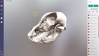
IVALA® - 3D Veterinary Anatomy - Promo
3 years agoUPD Canine skull - individual skull bones & sinus system - 3D Veterinary Anatomy & Learning IVALA®
87 -
 0:25
0:25
TheLearningDiary
4 months agoHuman Organ Facts #bodyfacts #generalknowledge #humanbodyfacts #healtheducation #shorts #shortvideo
43 -
 9:16
9:16
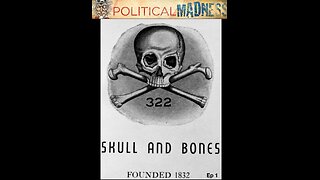
Political Madness
5 months agoEpisode 1 - Numerology in Media Headlines - 322 Skull & Bones
41 -
 8:54
8:54
Allen Young
11 months agoNames of all the bones in human body
8 -
 2:07
2:07
Video Creator
11 months agoHow many bones are in the human body ?
10 -
 1:04
1:04
IVALA® - 3D Veterinary Anatomy - Promo
3 years ago $0.08 earnedCalf - cow - bovine forelimb bone anatomy - 3D Veterinary Anatomy & Learning IVALA
6.77K -
 0:27
0:27
nodirjon
1 year agoDRAW A SKULL || draw a skull 💀💀💀👌✔
28