Myelopathy Patient Walks Again after Lumbar Surgery
Meet Juan.
He injured his back at work resulting in losing feeling in the nerves of his legs. Due to the damaged discs from this injury, he had problems feeling his toes and walking became extremely difficult. At Synapse Orthopedic Group, Edwin Haronian MD was able to diagnose him with Myelopathy. Haronian performed lumbar surgery on Juan and only two weeks after surgery, Juan was walking and feeling his nerves again.
Call us to schedule an appointment: (818) 788-2400
Locations in Pomona, Sherman Oaks, and Los Angeles
Disk herniation is a breakdown of a fibrous cartilage material (annulus fibrosis) that makes up the intervertebral disk. The annulus fibrosis surrounds a soft gel-like substance in the center of the disk called the nucleus pulposus. Pressure from the vertebrae above and below may cause the nucleus pulposus to be forced against the sides of the annulus. The constant pressure of the nucleus against the sides of the annulus will cause the fibers of the annulus to break down. As the fibers of the annulus break down, the nucleus will push toward the outside of the annulus and cause the disk to bulge in the direction of the pressure. This condition most frequently occurs in the lumbar region and is also commonly called a herniated nucleus pulposus, prolapsed disk, ruptured disk, or a slipped disk.
The spinal column is made up of 24 vertebrae that are joined together and permit forward and backward bending, side bending, and rotation of the spine. There are seven cervical (neck), twelve thoracic (chest region), and five lumbar (low back) vertebra. There are intervertebral disks between each of the 24 vertebrae as well as a disk between the lowest lumbar vertebrae and the large bone at the base of the spine called the sacrum.
Disk herniation most commonly affects the lumbar region. However, disk herniation can also occur in the cervical spine. The incidence of cervical disk herniation is most common between the fifth and sixth cervical vertebrae. The second most common area for cervical disk herniation occurs between the sixth and seventh cervical vertebrae. Disk herniation is uncommon in the thoracic region.
The peak age for occurrence of disk herniation is between 20 and 45 years of age. Studies have shown that males are more commonly affected than females in lumbar disk herniation by a 3:2 ratio. Long periods of sitting or a bent-forward work posture may lead to an increased incidence of disk herniation.
There are four classifications of disk pathology:
A protrusion occurs when a disk bulges without rupturing the annulus fibrosis.
A prolapse occurs when the nucleus pulposus pushes to the outermost fibers of the annulus fibrosis but does not break through them.
An extrusion occurs when the outermost layer of the annulus fibrosis is torn and the material of the nucleus moves into the epidural space.
A sequestration occurs when fragments from the annulus fibrosis or the nucleus pulposus have broken free and lie outside the confines of the disk.
Any direct or, forceful in a vertical direction pressure on the disks can cause the disk to push its nucleus into the fibers of the annulus or into the intervertebral canal. A herniated disk may occur suddenly from lifting, twisting, or direct injury, but more often it will occur from constant compressive loads over time. There may be a single incident that causes symptoms to be felt, but very often the disk was already damaged and bulging prior to any one particular incident.
Depending on the location of the herniation, the herniated material can also press directly on nerve roots or on the spinal cord. Pressure on the nerve roots or spinal cord may cause a shock-like pain sensation down the arms if the herniation is in the cervical vertebrae or down the legs if the herniation is in the lumbar region.
In the lumbar region a herniation that presses on the nerve roots or the spinal cord may also cause weakness, numbness, or problems with bowels, bladder, or sexual function. It is unclear if a herniated disk causes pain by itself without pressing on neurological structures. It is likely that irritation of the disk or the adjacent nerve roots may cause muscle spasm and pain in the region of the disk pathology.
Several radiographic tests are useful for confirming a diagnosis of disk herniation and locating the source of pain. X rays show structural changes of the lumbar spine. Myelography is a special x ray of the spine in which a dye or air is injected into the patient's spinal canal. The patient lies strapped to a table as the table tilts in various directions and spot x rays are taken. X rays showing a narrowed dye column in the intervertebral disk area indicate possible disk herniation.
For more information please visit our website at: http://www.synapsedoctor.com
You can also follow our social media pages at:
http://www.facebook.com/synapsedoctor
http://www.instagram.com/synapsedoctor
http://www.twitter.com/synapsedoctor
-
 7:30
7:30
TylerLebel
2 years agoHOW TO TAKE 40% LOAD OFF OF THE LUMBAR SPINE!! (PT2/6: LIFTING)
68 -
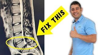 17:21
17:21
Bob The Physio
10 months agoLumbar Disc Bulges l4 l5 - l5 S1 recovery without surgery
28 -
 41:31
41:31
Bob The Physio
8 months agoHow to recover from lumbar disc extrusion at home and avoid surgery
1051 -
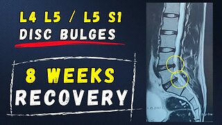 30:12
30:12
Bob The Physio
9 months agoL4 L5 L5 S1 Lumbar Disc bulge recovery at home
54 -
 23:53
23:53
Bob The Physio
1 year agoLumbar Disc Bulges L5 S1 recovery without surgery
51 -
 29:49
29:49
Bob The Physio
8 months ago $0.01 earnedHow to recover from lumbar l4 l5 / l5 S1 disc bulges at home
63 -
 16:29
16:29
Bob The Physio
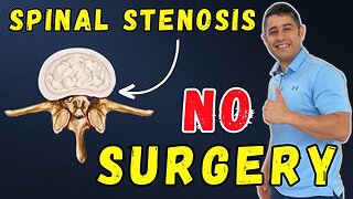
1 year agoChronic Lower Back pain due to spinal stenosis getting pain free without surgery
42 -
 0:51
0:51
Bob The Physio
1 year agoLumbar Spinal Stenosis
38 -
 40:47
40:47
Bob The Physio
1 year agoHow to recover from Lumbar Spine disc bulges L4 L5 L5 S1
47 -
 14:10
14:10
PerformancePlaceSportsCare
1 year agoSciatica: Is Sitting or Walking Worse For Your Leg Pain?
224