What are Haploid and Diploid Cells?
For Employees of hospitals, schools, universities and libraries: download up to 8 FREE medical animations from Nucleus by signing up for a free trial at: http://nmal.nucleusmedicalmedia.com/biology_youtube
#HaploidCells #DiploidCells #biology
SCIENCE ANIMATION TRANSCRIPT: In this video, we will discuss haploid versus diploid cells. Haploid and diploid are terms that describe the number of sets of chromosomes in a cell. Haploid means the cell has only one set of chromosomes. And diploid means the cell contains two sets of chromosomes. In your body, sex cells called Gametes have a haploid number of chromosomes represented by symbol n. In humans, every gamete has one set of 23 chromosomes, so the haploid, or n, number in humans is 23. This is important, since the union of gametes during fertilization creates a diploid cell called a zygote with two sets of chromosomes for a total of 46. At fertilization, the chromosomes from each parent match up to become the new pairs of chromosomes in a zygote. Each pair contains one chromosome from the father and a corresponding chromosome from the mother. These pairs are called homologous chromosomes. Homologous chromosomes are similar in shape and size along with the same types of genes in the same locations. A diploid zygote will go through cell division many times to produce all the cells in the body of a fully developed baby. All body cells except gametes are referred to as somatic cells. In humans, somatic cells are always diploid, written as 2n, which means they have 2 sets of 23 chromosomes for a total of 46 chromosomes. Other organisms have somatic cells with different diploid numbers of chromosomes. But the gametes in these organisms are haploid, meaning they always have half the diploid number of chromosomes. So, how does cell division affect the number of chromosomes in daughter cells? Well, somatic cells only reproduce by mitosis, a type of cell division that results in two genetically identical diploid daughter cells. In contrast, meiosis is a type of cell division that only produces gametes. In meiosis, a diploid cell undergoes two cell divisions to produce four genetically different haploid gametes. We'll cover the details of meiosis in another video. In summary, diploid cells have two complete sets of chromosomes. One set from each parent. Diploid cells have twice the number of chromosomes as haploid cells. The two sets consist of pairs of homologous chromosomes. The diploid chromosome number is written as 2n. All somatic cells, whether they're skin cells, muscle cells, or leaf cells in a plant are diploid. Diploid cells reproduce only by mitosis. And gametes are never diploid. In contrast, gamete cells, which are always haploid, have only one set of chromosomes, which is half the diploid number. Since there's only one set of chromosomes there are no homologous pairs. The haploid chromosome number is written as n. All gametes are haploid. And haploid gametes form from diploid cells through meiosis, never through mitosis. [music]
NSV15017
31
views
M Phase of the Cell Cycle
For Employees of hospitals, schools, universities and libraries: download up to 8 FREE medical animations from Nucleus by signing up for a free trial at: http://nmal.nucleusmedicalmedia.com/biology_youtube
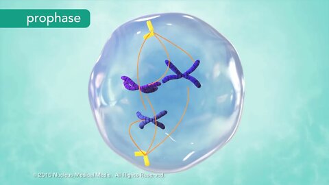
SCIENCE ANIMATION TRANSCRIPT: In this lesson, we'll be exploring the M phase of the cell cycle including mitosis and cytokinesis. Let's do a quick review of the cell cycle to see where they fit in. The G1, S, and G2 phases make up interphase, and the M phase represents cell division. Cell division includes division of the nucleus, called mitosis, and division of the cytoplasm, called cytokinesis. Mitosis is further broken down into four phases: prophase, metaphase, anaphase, and telophase. Prophase is the longest phase of mitosis. Prophase is when chromatin begins to condense into the shape of chromosomes, and the nucleolus disappears. The previously replicated DNA coils tightly into sister chromatids. For the first time, you see individual chromosomes. In the center of each chromosome, a centromere attaches the sister chromatids together. Meanwhile, in the cytoplasm, microtubules known as spindle fibers begin to fan out from two sets of paired structures called centrioles. The spindle fibers elongate as the centrioles begin moving to opposite sides, or poles, of the cell. While this is happening, the nuclear membrane surrounding the nucleus disappears. Now that chromosomes are no longer separated from the cytoplasm, the opposite ends of the spindle fibers can attach to the centromeres. Next, the cell enters metaphase. The centrioles complete their movement to the poles of the cell while the spindle fibers line up the chromosomes along the equator of the cell. The end-to-end alignment of chromosomes results in a sister chromatid on either side of the equator. Anaphase follows metaphase. During anaphase, spindle fibers separate the sister chromatids at their centromere. Once separated from each other, each chromatid is called a chromosome. The single-stranded chromosomes form a V shape as the spindle fibers shorten and drag them through the gel-like cytoplasm. The chromosomes move to opposite poles of the cell toward their centrioles. It's common to confuse centrioles with centromeres which connect chromatids. Remember, centrioles are at the poles. Telophase is the final stage of mitosis. In telophase, a nuclear membrane re-forms around each set of chromosomes. Then the chromosomes spread out into chromatin, and the nucleolus becomes visible once again. Mitosis, the division of the nucleus, is now complete. The final step of the M phase is cytokinesis, the division of the cytoplasm. In animal cells, cytokinesis occurs through the inward movement of the cell membrane. This progressively pinches the cytoplasm until two identical daughter cells form. In contrast, plant cells can't pinch in two because they have a rigid cell wall surrounding their cell membrane. Instead, cell wall material assembles along the equator forming a structure called the cell plate. The cell plate grows until it joins with the existing cell membrane, separating the two halves of the cell into daughter cells. Over time, new cell walls form between the two daughter cells. Here are the key points to remember. The M phase is the fourth and final phase of the cell cycle. During the M phase, cell division occurs through two processes: mitosis, when the nucleus divides, and cytokinesis, when the cytoplasm divides. Mitosis has four phases. During prophase, chromatin condenses into chromosomes, spindle fibers form, and the nucleolus and nuclear membrane disappear. During metaphase, spindle fibers align the chromosomes along the cell equator. In anaphase, the spindle fibers separate sister chromatids into two separate groups of chromosomes, pulling them toward the poles. And in telophase, the nucleolus and nuclear membrane re-form. The chromosomes disperse into chromatin. Cytokinesis is division of the cytoplasm. The M phase is complete after cytokinesis occurs. The M phase of the cell cycle always results in two daughter cells. Both of these daughter cells are identical to each other and identical to the original cell that underwent mitosis. [music]
NSV15006
38
views
The Cell Cycle
For Employees of hospitals, schools, universities and libraries: download up to 8 FREE medical animations from Nucleus by signing up for a free trial at: http://nmal.nucleusmedicalmedia.com/biology_youtube
SCIENCE ANIMATION TRANSCRIPT: In this lesson, we'll be looking at the cell cycle. This is the lifespan of a eukaryotic somatic cell. A somatic cell is any cell in the body of an organism, except for sex cells such as sperm and egg cells. The cell cycle describes the sequence of cell growth and division. A cell spends most of its life a state called interphase. Interphase has three phases, the G1, S, and G2 phases. Interphase is followed by cell division, which has one phase, the M phase. Together these four phases make up the entire cell cycle. G1 of interphase is sometimes called growth 1 or gap phase 1. In G1, a cell is busy growing and carrying out whatever function it's supposed to do. Note that some cells, such as muscle and nerve cells, exit the cell cycle after G1 because they do not divide again. A cell enters the S phase after it grows to the point where it's no longer able to function well and needs to divide. The S stands for synthesis, which means to make, because a copy of DNA is being made during this phase. Once DNA replication is complete, the cell enters the shortest and the last part of interphase called G2, also known as growth 2 or gap phase 2. Right now, it's enough to know that further preparations for cell division take place in the G2 phase. Now that interphase is over, the cell is ready for cell division, which happens in the M phase. The M phase has two events. The main one is mitosis, which is division of the cell's nucleus, followed by cytokinesis, a division of the cytoplasm. So, at the end of M phase, you have two daughter cells identical to each other and identical to the original cell. Let's review. The cell cycle describes the life cycle of an individual cell. It has four phases, three in interphase and one for cell division. Most cell growth and function happen during G1. The cell enters the S phase when it needs to divide. In this phase the cell replicates its DNA. Replication just means the cell makes a copy of its DNA. In G2, the cell undergoes further preparations for cell division. Finally, we have cell division in the M phase. The M phase consists of mitosis, which is nuclear division, and cytokinesis, or division of the cytoplasm. We'll explore the details of mitosis and cytokinesis separately. [music]
NSV15004
38
views

Overview of Cell Division
For Employees of hospitals, schools, universities and libraries: download up to 8 FREE medical animations from Nucleus by signing up for a free trial at: http://nmal.nucleusmedicalmedia.com/biology_youtube
SCIENCE ANIMATION TRANSCRIPT: In this lesson, we'll be talking about how cells reproduce. How and why do they do this? Well, they use a process called cell division to make new cells called daughter cells. Unicellular organisms, meaning creatures that consist of just one cell such as bacteria, usually clone themselves during cell division. The two daughter cells that result are separate organisms, in this case, two new genetically identical bacteria. This is a type of asexual reproduction known as binary fission. Cells in a multicellular organism also reproduce by cell division, but the new daughter cells that are produced are not two separate organisms. Instead, these new cells are just parts of the organism, allowing it to grow, or sometimes replacing cells that are worn out or injured. For example, your body heals a paper cut through division of your skin cells, occurring at the edges of the cut. In a modified example of cell division, sex cells called gametes are made. Chromosomes are an important part of cell division. So, what are chromosomes? Let's look inside a cell's nucleus. Here we find the nuclear genetic material known as deoxyribonucleic acid or DNA. Each cell's DNA holds the genetic code or instructions from everything within that organism. Looking through a microscope, you can see that DNA is usually spread out within the nucleus. It looks kind of grainy. We call the DNA Chromatin when it looks like this. Before a cell divides, DNA must replicate or copy itself so that the information in this code can be passed on to each daughter cell. At the beginning of cell division, DNA condenses tightly into an x-shaped structure known as a chromosome. Each side of an x-shape chromosome is a genetically identical sister chromatid, forming a sideways v-shape. In the middle, a structure called a centromere, joins the sister chromatids together. Different types of organisms have different numbers of chromosomes. Every cell in your body is called a somatic cell, except your gametes. Human somatic cells have 23 pairs of chromosomes, for a total of 46. It's like the difference between how many pairs of shoes you have versus your total number of shoes. Gametes are the exceptions to this rule. Human eggs and sperm have only one chromosome from each pair, for a total of 23. And unlike somatic cells, gametes are not genetically identical to their parent cells. When a sperm cell fertilizes an egg, they both contribute their 23 chromosomes. This fused cell, called a zygote, now has 23 pairs of chromosomes for a total of 46. This overview of cell division will help prepare you for studying the cell cycle, which is the life cycle of the cell. [music]
NSV15003
50
views
Cell Transport and Solutions
For Employees of hospitals, schools, universities and libraries: download up to 8 FREE medical animations from Nucleus by signing up for a free trial at: http://nmal.nucleusmedicalmedia.com/biology_youtube
#CellTransport #CellSolutions #biology
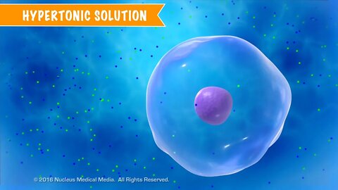
SCIENCE ANIMATION TRANSCRIPT: In this video, we'll discuss cell transport and solutions. The concentration gradient of extracellular solutions affects the transport of substances through the cell membrane. What can happen in this process? Well, cells may be surrounded by solutions with different particle concentrations, both in a lab as well as inside a living organism. So we need to know how cells will respond. Compared to the solution inside a cell, a solution outside the cell may be hypertonic, isotonic, or hypotonic. We'll describe hypertonic solutions using this beaker of water with dissolved salt, and a rather large cell submerged in it. To demonstrate water movement in and out of cells, we'll use simple numbers to illustrate concepts. Remember, the cell cytoplasm is mostly water as well. In this example, let's say the cytoplasm inside the cell is a solution containing 10% salt, which means the other 90% is water. Keep in mind percentages of substances within the cell must equal 100%. The percentages of solute and solvent in the beaker must also add up to 100%. However, the solution in the beaker is a different concentration from the solution inside the cell. It's 20% salt and 80% water. So you can see that there's a greater concentration of water inside the cell than outside, 90% compared to 80%. We call a solution hypertonic when its concentration of water is lower than inside the cell and its concentration of solute is higher than inside the cell. As a result, water molecules diffuse out of the cell through osmosis, causing the cell to shrivel a bit. Remember, osmosis means diffusion of water. If the solution's water concentration was significantly lower, the cell could shrivel to the point of imploding. For red blood cells in hypertonic solutions, the shriveling from osmotic water loss is called crenation. Let's look at an example of plant cells in a hypertonic solution. Normally, turgor pressure, which is the water pressure in a plant cell's central vacuole, helps support the cell wall and overall plant shape. When water leaves plant cells by osmosis, the cell membrane and its contents shrink away from the rigid cell wall, and turgor pressure decreases. This is called plasmolysis. Plasmolysis causes a plant to wilt. Let's look at another container, this time with a cell floating in an isotonic solution. Sometimes, the solution outside the cell has about the same concentration gradient as the concentration gradient inside the cell. Once again, in this example, the solution inside the cell is 90% water with 10% salt, but this time, the surrounding solution is also 90% water with 10% salt dissolved. We call a solution isotonic when its concentrations of water and solute are the same as inside the cell. But this doesn't mean that there is no movement. In an isotonic solution, the rate of water molecules entering the cell is equal to the rate of water molecules exiting the cell. The amount of water molecules going in equals the amount of water molecules going out. That's why you see the arrows pointing in two different directions. In an isotonic solution, the cell doesn't shrink or swell. It stays exactly the same size. A third possibility is a solution outside the cell with a higher concentration of water than the solution inside the cell. In this example, the solution inside the cell has 20% salt. So that means the remainder is 80% water, but the cell has been placed in a beaker that only has a 10% salt solution. Doing the math, we can see that the solution surrounding the cell is 90% water, which means there's a greater concentration of water outside the cell than inside. We call a solution hypotonic when its concentration of water is higher than inside the cell, and its concentration of solute is lower than inside the cell. So by osmosis, the water molecules will move passively into the cell until a state of equilibrium is reached. Hypotonic solutions cause a cell to swell up with water. If the water concentration outside the cell is high enough, the cell can swell to the point of bursting. This is called cytolysis. Cytolysis in red blood cells is called hemolysis. Cytolysis doesn't happen in plant cells because the rigid cell wall prevents the cells from bursting. Here's a little trick to remember that hypotonic solutions cause a cell to swell rather than shrivel. When you think of hypo, think of a big, swollen hippopotamus or hippo for short. So to recap, we can have solutions that are hypertonic, isotonic, or hypotonic with respect to the cell. In an isotonic solution, the water concentration inside and outside the cell stays about the same. The water concentration stays the same because the concentration of solute is the same...
NSV15018
116
views
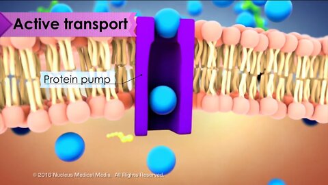
Cell Biology: Active Transport
For Employees of hospitals, schools, universities and libraries: download up to 8 FREE medical animations from Nucleus by signing up for a free trial at: http://nmal.nucleusmedicalmedia.com/biology_youtube
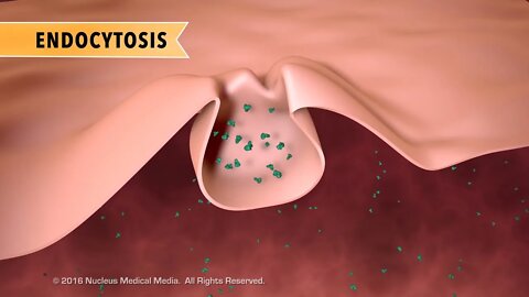
SCIENCE ANIMATION TRANSCRIPT: In this video, we'll discuss active transport. Active transport is when particles move from an area of low concentration to high concentration. This is also known as moving against the concentration gradient. The key thing to remember is that active transport requires energy. If passive transport is like a ball naturally rolling down a hill, active transport is the opposite. You can get the ball back up the hill, but you're going to have to expend some energy to do it. Cells require this type of substance movement in order to function properly. For example, heart muscle cells responsible for making your heart beat, move molecules or ions against their concentration gradient. So, what are some of the main types of active transport? We have endocytosis, exocytosis, and protein pumps. Sometimes a cell uses active transport to pull in large particles using its cell membrane. This is called endocytosis. One type of endocytosis is called phagocytosis. This often happens when the cell takes in some type of nutrient. In another type of endocytosis called pinocytosis, the cell takes in fluids by creating pockets in the cell membrane. The cell can ingest a large amount of fluid this way by pinching off these cell membrane pockets into the cytoplasm. The opposite of endocytosis is exocytosis. Exocytosis is when something needs to exit the cell. The cell can remove large molecules or wastes this way by fusing the membrane bound vesicles containing them with the cell membrane, forcing them out of the cell. A good way to remember that exocytosis is a way for things to leave the cell is that it shares the first two letters with exit. You can also remember that endocytosis is a way for things to move into the cell because each shares the first two letters with enter. Sometimes the cell uses special protein pumps to move small molecules or ions against the concentration gradient into or out of the cell. An example of this is the sodium potassium pump. In this process, the pump uses energy in the form of ATP molecules to move sodium ions out of the cell and then move potassium ions into the cell. Protein pumps used an active transport require energy because the molecules or ions are moving from an area of low concentration to high concentration. In summary, active transport is when the cell uses energy to move substances in or out of the cell against the concentration gradient via endocytosis, exocytosis, or protein pumps. [music]
NSV15009
9
views
Biology Quiz: What are organelles?
Tell us what you think about our new Biology Quiz shorts, and subscribe to our new Biology channel to get more quizzes and biology animations!
#shorts #biology #cells
6
views
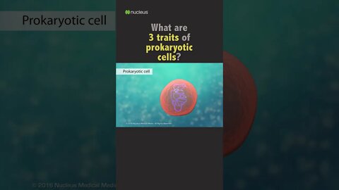
Biology Quiz: 3 traits of prokaryotic cells?
Tell us what you think about our new Biology Quiz shorts, and subscribe to our new Biology channel to get more quizzes and biology animations!
#biology #shorts #cells
4
views
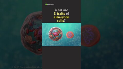
POP QUIZ: 3 traits of eukaryotic cells?
Tell us what you think about our new Biology Quiz shorts, and subscribe to the Nucleus Biology channel to get more quizzes and biology animations!
#shorts #biology #cells
1
view

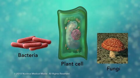
1 Minute Biology Quiz - 2 Categories of Cells
Subscribe to Nucleus Biology to prep for your next test! This #short is What are the 2 categories of cells?
#shorts #biology #quiz
1
view
Overview of Cell Structure
For Employees of hospitals, schools, universities and libraries: download up to 8 FREE medical animations from Nucleus by signing up for a free trial at: http://nmal.nucleusmedicalmedia.com/biology_youtube
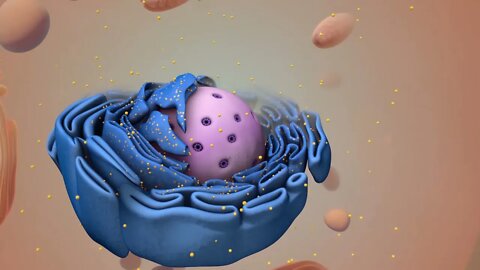
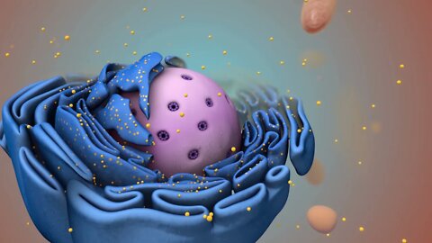
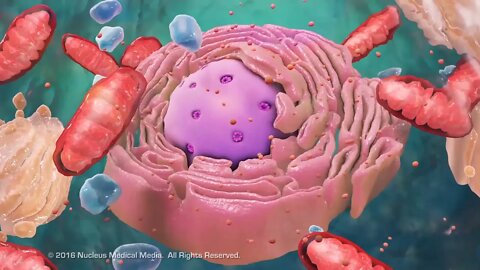
SCIENCE ANIMATION TRANSCRIPT: [music] Cells are the smallest living units of an organism. All cells have three things in common, no matter what type of cell they are. All cells have a cell membrane which separates the inside of the cell from its environment. Cytoplasm, which is a jelly-like fluid, and DNA, which is the cell's genetic material. There are two broad categories of cells. The first category is eukaryotic cells. They have organelles which include the nucleus and other special parts. Eukaryotic cells are more advanced complex cells such as those found in plants and animals. The second category is prokaryotic cells. They don't have a nucleus or membrane-enclosed organelles. They do have genetic material, but it's not contained within a nucleus. Prokaryotic cells are always one-celled or unicellular organisms, such as bacteria. [music] So, what are organelles? Organelle means little organ. Organelles are the specialized parts of a cell that have unique jobs to perform. Let's start with the nucleus, the control center of the cell. The nucleus contains DNA, or genetic material. DNA dictates what the cell is going to do and how it's going to do it. Chromatin is the tangled spread out form of DNA found inside the nuclear membrane. When a cell is ready to divide, DNA condenses into structures known as chromosomes. [music] The nucleus also contains a nucleolus, which is a structure where ribosomes are made. After ribosomes leave the nucleus, they will have the important job of synthesizing, or making, proteins. [music] Outside the nucleus, the ribosomes and the rest of the organelles float around in cytoplasm, which is the jelly-like substance. Ribosomes may wander freely within the cytoplasm or attach to the endoplasmic reticulum, sometimes abbreviated as ER. There are two types of ER. Rough ER has ribosomes attached to it. And smooth ER doesn't have ribosomes attached to it. The endoplasmic reticulum is a membrane-enclosed passageway for transporting materials such as the protein synthesized by ribosomes. Proteins and other materials emerge from the endoplasmic reticulum in small vesicles where the Golgi apparatus, sometimes called the Golgi body, receives them. As proteins move through the Golgi body, they are customized into forms that the cell can use. The Golgi body does this by folding the proteins into useable shapes or adding other materials onto them such as lipids or carbohydrates. Vacuoles are sack-like structures that store different materials. Here in this plant cell, the central vacuole stores water. [music] Going back to the animal cell, you will see an organelle called a lysosome. Lysosomes are the garbage collectors that take in damaged or worn out cell parts. They are filled with enzymes that break down the cellular debris. The mitochondrion is an organelle that is the powerhouse for both animal and plant cells. During a process called cellular respiration, the mitochondria make ATP molecules that provide the energy for all of the cells activities. Cells that need more energy have more mitochondria. [music] Meanwhile, the cell maintains its shape through a cytoskeleton. The cytoskeleton includes the thread-like microfilaments which are made of protein, and microtubules which are thin, hollow tubes. Some organisms such as plants that are photoautotrophic, meaning they capture sunlight for energy, have cells with an organelle called a chloroplast. The chloroplast is where photosynthesis happens. It's green because it has a green pigment called chlorophyll. Plant cells also have a cell wall outside of their cell membranes that shape, support, and protect the plant cell. Animal cells never have a cell wall. There are many other unique structures that only some cells have. Here are just a few. In humans, for example, the respiratory tract is lined with cells that have cilia. These are microscopic, hair-like projections that can move in waves. This feature helps trap inhaled particles in the air and expels them when you cough. Another unique feature in some cells is flagella. Some bacteria have flagella. A flagellum is like a little tail that can help a cell move or propel itself. The only human cell that has a flagellum is a sperm cell. In summary, remember, eukaryotic cells are plant and animal cells with a nucleus and membrane-enclosed organelles. While prokaryotic cells are unicellular organisms without these things. All cells have a cell membrane, cytoplasm, and genetic material. And even though only plant cells have chloroplast, both plant and animal cells have mitochondria. [music]
NSV15001
66
views
Overview of Cell Boundaries
For Employees of hospitals, schools, universities and libraries: download up to 8 FREE medical animations from Nucleus by signing up for a free trial at: http://nmal.nucleusmedicalmedia.com/biology_youtube
SCIENCE ANIMATION TRANSCRIPT: Today, we're going to talk about the outer boundary of cells. Every cell has a boundary to separate it from its surroundings. You may already know that plant cells have a rigid outer boundary called a cell wall. Other organisms such as bacteria and fungi also have cell walls. And while their cell walls differ in structure and composition, their cell walls all provide support, shape, and protection for these types of cells. It is essential you remember that animal cells always have a cell membrane but never have a cell wall. So what is the boundary that all cells have? Whether they have a cell wall or not, all cells have a cell membrane, also called a plasma membrane. In a typical animal cell, the cell membrane is a thin, flexible barrier against the outside environment. It's main job is to help with homeostasis, a type of equilibrium in which the cell maintains a relatively constant, stable internal environment. Like all living things, cells require stable internal conditions in order to survive, grow, and reproduce. The cell membrane helps maintain this stable internal environment by being selectively permeable. This means it acts as a gatekeeper to control or select what can get into or out of the cell. We'll learn more about the ways cells accomplish this separately. For now, remember that all cells have a flexible cell membrane, and most cells also have a rigid cell wall. And it's important to know that animal cells never have a cell wall. The cell wall provides support, shape, and protection to the cell. And the cell membrane is selectively permeable in order to help maintain intracellular homeostasis. [music]
NSV15002
12
views
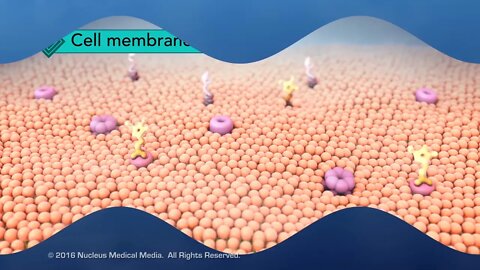
Structure of the Cell Membrane
For Employees of hospitals, schools, universities and libraries: download up to 8 FREE medical animations from Nucleus by signing up for a free trial at: http://nmal.nucleusmedicalmedia.com/biology_youtube
SCIENCE ANIMATION TRANSCRIPT: In this video, we will be discussing the structure of the cell membrane. When scientists looked at the selectively permeable cell membrane, they described its structure as a fluid mosaic. You might know that a mosaic is a picture made up of little tiles. Like a mosaic, the cell membrane is made up of different parts as well. The cell membrane has two layers of phospholipids referred to as a lipid bilayer. The lipid bilayer isn't rigid. The phospholipids in it have the ability to move in a flexible wave-like motion. Let's take a closer look at a few phospholipids. The round head portions are hydrophilic, which means they're attracted to water. Both the extracellular fluid, meaning fluid outside the cell, and the cytoplasm inside the cell are mostly made up of water. So, the hydrophilic phospholipid heads of the outer layer will be oriented toward the extracellular fluid. And the heads of the inner layer will be oriented toward the cytoplasm. The phospholipid tails are hydrophobic, which means watery areas withheld them. So they orient toward each other in a direction as far away from the watery content as possible. There are also scattered proteins embedded in the phospholipid layers, some with carbohydrates attached. So, in the fluid mosaic model, the cell membrane is made up of different parts. And these parts make up a flexible boundary around the cell. But how do the majority of substances get in our out of the cell? Some molecules sip through the little spaces in between the phospholipids, which make up the majority of the semi-permeable cell membrane. However, other molecules are too big to fit through the cell membrane this way. So, how do these larger molecules pass through the cell membrane? The molecules move through proteins embedded in the cell membrane, either from the extracellular area into the cell, or from the intracellular area out of the cell. These substances will move through tunnels made up of these proteins. We'll explore how things move through the cell membrane in greater detail separately. [music]
NSV15005
9
views
Overview of Cell Transport
For Employees of hospitals, schools, universities and libraries: download up to 8 FREE medical animations from Nucleus by signing up for a free trial at: http://nmal.nucleusmedicalmedia.com/biology_youtube
SCIENCE ANIMATION TRANSCRIPT: Cell transport is the process of how things move in or out of the cell through the cell membrane. There are two broad categories of cell transport. The first category is passive transport. For a cell, passive transport means it's an automatic process that doesn't require any input of energy. For example, diffusion is a passive process in which particles move either into or out of the cell from an area of higher concentration to an area of lower concentration. The cell doesn't use any energy when this happens. The second category of cell transport is active transport. This is when particles move from an area of lower concentration to an area of higher concentration. When particles move against the concentration gradient, energy is required often to allow protein pumps to assist in particle movement. Why would the cell need to move particles from a low to high concentration and expend energy to do it? An important example is seen in your heart muscle cells. In order for your heart to beat, there are certain molecules that have to move from an area of low concentration to an area of high concentration for those cardiac muscle cells to work. So, the main things to remember are passive transport happens automatically with no energy required, while active transport needs energy for it to occur. [music]
NSV15007
4
views
PassiveTransport
For Employees of hospitals, schools, universities and libraries: download up to 8 FREE medical animations from Nucleus by signing up for a free trial at: http://nmal.nucleusmedicalmedia.com/biology_youtube
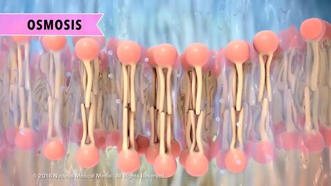
SCIENCE ANIMATION TRANSCRIPT: In this video, we will be discussing passive transport. Passive transport is when particles move through the cell membrane from an area of higher concentration to lower concentration without the use of energy, also described as movement along the concentration gradient. What are the types of passive transport? They are diffusion, osmosis, and facilitated diffusion. We'll describe diffusion first using the solution in this container. A solution is a liquid with something dissolved in it. The aqua color represents the solvent, meaning the liquid part of the solution. The yellow particles represent the dissolved substance called the solute. The structure in the middle of the container represents a semi-permeable cell membrane, a barrier through which only certain sized particles can pass freely. It's important to note that although diffusion often occurs across the cell membrane, diffusion can happen with or without a semi-permeable membrane. Right now, there is more solute on the left than there is on the right. Because solute particles are able to pass through the semi-permeable membrane, they are going to naturally move from an area of high concentration to an area of low concentration. They will continue to do this until both sides of the container have about equal numbers of solute particles. This is called achieving a state of equilibrium. Let's review what we've covered so far. Diffusion is when particles move from an area of high concentration to low concentration. This just happens. It's a natural process that doesn't use any energy. Here's an example of diffusion happening without a semi-permeable membrane. If you spray air freshener in a room, people near you smell it right away. But after a short time, depending on the size of the room, people farther away will also begin to smell it. This is because the little scented molecules are trying to achieve equilibrium by spreading evenly throughout the room. Remember, diffusion is a natural process, like a ball rolling down a hill. The ball's movement is automatic and doesn't require any energy. Osmosis is diffusion that happens with water molecules. Let's look at another container in which the solvent is water but the solute particles are larger. The membrane in this container has openings that are too small for the solute to move through, but water can pass through the membrane freely. This time, we'll focus on the concentration gradient of the water rather than the solute particles. Although the large solute particles can't pass through the membrane, the water molecules are small enough to pass through. The water moves freely from its area of high concentration to low concentration until equilibrium is reached. Equilibrium means that the proportion of water to solute particles is about the same on both sides of the membrane. In the cell, osmosis means diffusion of water through the cell membrane. Water can enter or leave the cell through the membrane until the cell achieves a state of equilibrium with its surroundings. So like diffusion, osmosis is passive. No energy is required. It just happens automatically. Facilitated diffusion is a type of passive transport in which molecules diffuse through specialized protein channels in the cell membrane. The protein channels work like special ports or tunnels that allow these substances in or out of the cell. Facilitated diffusion is also when particles move from high concentration to low concentration. How do you know that? From the word "diffusion." Facilitated diffusion works naturally without added energy, just like the diffusion example we discussed earlier. But facilitated diffusion generally happens with particles a bit larger than those that can seep through the cell membrane's phospholipid layers. So they move in or out of the cell along the concentration gradient in a specialized way through protein channels. In summary, passive transport is a natural process that doesn't require the cell to expend any energy. The types of passive transport are diffusion, osmosis, and facilitated diffusion. [music]
NSV15008
35
views
1 Minute Biology Quiz - 3 things all cells have in common?
This video is a #short flash card quiz on cell biology with a quick review of the material afterward. Question: What are three things all cells have in common?
#biology #shorts #cells
3
views